Retinal Vein Occlusion
What is Retinal Vein Occlusion?
In the eye (as well as all other body tissues except the lungs), arteries deliver blood to a tissue and veins remove blood from a tissue. The arteries carry oxygen and nutrition to the tissue, while veins remove waste products. A tiny retinal vein can become blocked by a blood clot; this blockage creates a backup in the system, resulting in leakage of blood and plasma (fluid from the bloodstream) into the retinal tissue. This is a retinal vein occlusion. The retinal bleeding (hemorrhage) and swelling (edema) can cause significant visual loss. In addition, disruption of the blood supply can cause permanent damage to the retinal tissue from lack of oxygen and nutrition. In some cases, this may result in the development of abnormal blood vessels as a response to the lack of oxygen.
This condition typically occurs in individuals greater than 50 years old. Risk factors include hypertension, cardiovascular disease, diabetes, and glaucoma. Sometimes, individuals with retinal vein occlusion are found to have blood clotting disorders or inflammatory conditions. The second eye is affected in approximately 10% of cases.
Get in touch
Retinal Vein Occlusion Form
We will get back to you as soon as possible.
Please try again later.
Retinal Vein Occlusion Symptoms
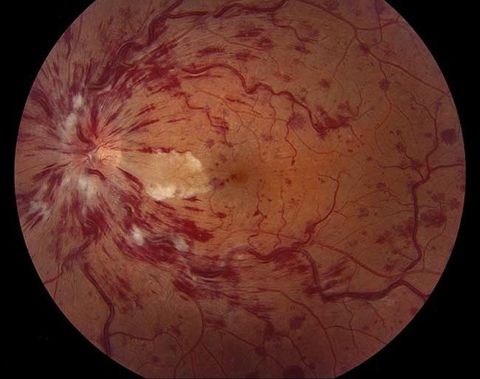
Retinal vein occlusions are classified as central retinal vein occlusion (CRVO) if the blockage occurs in the main vein leaving the eye through the optic nerve They are classified as branch retinal vein occlusion (BRVO) if the blockage occurs at one of the branches before reaching the main vein at the nerve; these often occur where arteries and veins cross. When patients have a retinal vein occlusion, they frequently complain of blurred vision, distortion, and/or a central blind spot.
Occluded veins typically appear tortuous, just as a hose tends to twist when water flow is blocked. There is bleeding seen in the distribution of the blocked vein in BRVO; the whole retina is involved in CRVO. There is also usually swelling of the retina in the same distribution; macular edema is a common finding and is often a source of significant visual loss. When there is a severe blockage in which blood flow is cut off or significantly reduced, sensitive retinal tissue can become permanently damaged. This process is called ischemia. As a response to ischemia, the eye may develop abnormal new blood vessels called neovascularization. Unfortunately, these new vessels are fragile and tend to bleed, potentially resulting in vitreous hemorrhage. They may also scar and pull on the retina, potentially resulting in traction retinal detachment. Finally, if these blood vessels grow in the drainage structure of the eye, a very serious form of glaucoma can develop called neovascular glaucoma.
Retinal Vein Occlusion Treatment
Treatment of retinal vein occlusion is aimed at macular edema and complications of neovascularization. The most effective strategy involves reducing the effect from external risk factors such as glaucoma, hypertension and dyslipidemia. In addition, retinal laser and Anti-Vegf injections can also reduce the swelling of the retina and prevent new blood vessel growth that can damage the eye severely.
Call
Office: 780-424-2233
Fax: 780-426-7219
All Rights Reserved | Visionary Eye Surgeons | Website Created by CCC
